For the first time, inflammatory processes in joints can be shown in 3D using molecular imaging techniques
We still do not know exactly what causes chronic inflammatory joint diseases such as rheumatoid arthritis. A team at the Department of Medicine 3, led by Prof. Gerhard Krönke, has now chosen a new approach to gain a better understanding of the underlying mechanisms. Using technology they have developed themselves, Stephan Culemann and Dr. Anika Grüneboom have succeeded for the first time in making inflamed joints transparent, allowing them to be investigated using three dimensional molecular imaging. The insights thus gained into the complex working of our immune system were published in the magazine ‘Nature’.
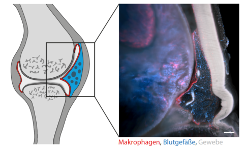
It was shown that healthy joints are coated in a continually renewing membrane of special immune cells (macrophages). Whilst this barrier of macrophages usually prevents our joints from being attacked by our own immune system, this protective mechanism fails in the event of rheumatoid arthritis. As a result, falsely activated immune cells can suddenly enter, leading eventually to the inflammation and destruction of the joint. Whilst macrophages have been suspected of contributing to joint inflammation, the current analyses show that they actually create an important anti-inflammatory protective sheath around the joint and are capable of curbing inflammatory reactions.
Further information
Prof. Dr. Gerhard Krönke
Phone: +49 9131 85 34742